Contrast enhanced mammography (CEM) is an exciting way to image the breast. Mammography significantly reduces mortality from breast cancer as a result of early detection. However, it can be difficult to detect breast cancer in women with very dense breast tissue. The ratio of fatty tissue to breast tissue is what determines breast density. New screening and diagnostic methods are being devised in order to overcome the difficulty of imaging dense breasts.
One of these methods is contrast enhanced mammography (CEM), also called contrast enhanced spectral mammography (CESM). The use of an intravenous contrast dye and digital mammography as used with CEM can increase the breast cancer detection rate. Standard screening mammograms can miss breast cancers due to their inability to “see through” dense breast tissue.
What is Contrast Enhanced Mammography (CEM)?
The FDA approved contrast enhanced mammography in 2011. It is widely used around the world and is gaining popularity – albeit more slowly – in the United States. CEM is a special mammogram that uses an intravenous iodine based contrast dye. CT scans use the same dye. A breast cancer needs to develop its own blood supply in order to grow. This is called angiogenesis. The contrast dye makes it easier to see cancers due to the increased blood flow to the cancer. These new blood vessels preferentially take up the contrast because of increased flow. In this regard, contrast enhanced mammography is similar to magnetic resonance imaging (MRI) of the breast.
How is Contrast Enhance Mammography (CEM) Performed?
An IV is started in the arm immediately before the mammogram. A digital mammogram is performed 2 minutes after the IV contrast is infused. The exam takes about 5 to 10 minutes and will feel just like a regular mammogram.
The image that CEM creates is a composite of two images taken at the same time – one showing the contrast and the other a regular mammogram. A computer program “subtracts” the images and creates an “iodine” or contrast only image which shows the areas that have contrast enhancement. Cancers usually take up more contrast than the normal breast tissue and will show up as “white” while the normal breast tissue will show up mostly dark.
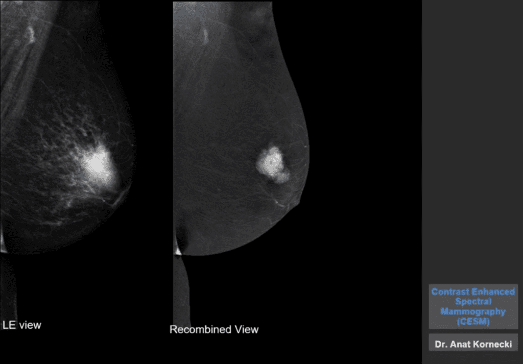
The following example has been provided by courtesy of Dr. Anat Kornecki. It shows a regular mammogram on the left and the CEM on the right. The regular mammogram shows a very dense area of breast tissue. However, we don’t see a specific cancer within the density. The image on the right shows the “subtraction” image where all the dense breast tissue disappears and the cancer is seen very clearly.
Indications for Contrast Enhanced Mammography
One of the most common uses of CEM in newly diagnosed women is to evaluate the extent of the breast cancer. This is particularly important in women with dense breast tissue. With CEM we can determine the size of the tumor and whether there are other abnormal areas in either breast. This allows for more accurate surgical planning. Many patients with triple negative breast cancer receive chemotherapy before surgery. The response to neo-adjuvant chemotherapy can be assessed with CEM. It can also be assessed by MRI. This topic has been covered in a previous Expert Forum post by Dr. Sarah Dodge.
It can also help problem solve when there is a question of recurrence at a lumpectomy site. It can help find hidden breast cancers when we know there are axillary lymph nodes metastases but can’t see the cancer on a regular mammogram. Patients who are not candidates for an MRI due to a pacemaker, or who cannot have an MRI due claustrophobia are excellent candidates for CEM.
Finally, your doctor may recommend that you have a CEM to look for breast cancer if the mammogram or ultrasound are inconclusive or if there are symptoms indicative of a possible cancer.
Advantages of Contrast Enhanced Mammography
CEM is almost equal to MRI in its ability to detect breast cancer and it is better than mammography and ultrasound combined. Contrast enhanced mammography is less expensive than MRI and takes about 10 minutes to perform compared to 30-40 minutes for MRI. CEM is particularly useful for patients with claustrophobia, leading to higher satisfaction among patients. In women at increased risk for breast cancer with dense breast tissue, CEM detects cancer more often than when compared to mammography alone.
Disadvantages of Contrast Enhanced Mammography
The drawbacks of CEM include the risk of contrast-induced allergic reactions. The radiation dose is slightly higher than a routine digital mammogram but still less than a 3D mammogram.
Injury to the kidneys may also result from exposure to iodinated contrast material if the patient has kidney disease. Assuring the patient has adequate renal function before CEM is a must. The use of CEM is contraindicated in patients with kidney disease or an allergy to the IV contrast.
Conclusion
Contrast enhanced mammography (CEM) is a valuable tool in the diagnosis and staging of primary triple negative breast cancer. It combines an iodinated contrast dye with conventional mammography to improve diagnostic accuracy, particularly in women with dense breast tissue.
MRI remains the method of choice, but when compared to CEM, CEM is cheaper, faster, and with similar sensitivity for the diagnosis of invasive breast cancer. The utilization of CEM in the United States is currently low but could increase rapidly given the many potential indications for its clinical use. You should speak to your healthcare provider to determine which method of detection and diagnosis is best for you.